Atlantoaxial Luxation
Back to Fact Sheets
The atlanto axial (AA) joint is the joint between the atlas and the axis, which are the first two vertebrae of the neck. Unlike the rest of intervertebral joints, in the atlantoaxial joint there is no intervertebral disc and the joint is entirely supported by ligaments.
Download PDF
Atlanto-axial luxation is a condition in which there is instability between the atlas and the axis. This is likely to happen secondary to a malformation in the axis (second vertebra) that is missing a very important part called the dens, which is fundamental for the AA joint. This disorder is more commonly seen in small and toy breed dogs like Chihuahuas, Pomeranians, Yorkshire Terriers and Miniature Poodles, but can affect big dogs and occasionally cats. In most cases, especially in predisposed breeds, this condition is related to a congenital abnormality, but sometimes can be caused by trauma.
What are the clinical signs of atlanto-axial luxation?
This movement between the atlas and the axis can cause pressure on the spinal cord and development of clinical signs. Commonly affected dogs show signs of neck pain, such as rigidity, crying or being reluctant to move the head. In more severe cases, compression of the spinal cord can lead to neurological deficits of all four legs, causing weakness or incoordination. In severe cases, a animal could be completely paralyzed or even die if the compression of the spine is really severe at this level.
Most of the dogs are young when they start to show clinical signs, but in some cases signs can start during adulthood, particularly if there is a trauma to that area.
Diagnose of atlanto-axial luxation?
To reach a diagnosis the patient needs to undergo a complete neurological exam, followed by imaging techniques to allow visualization of the abnormality. Radiography has been traditionally used for diagnosis, however this provides limited information about any bone deformity and no information regarding the severity of a possible spinal cord injury. This is the reason why advanced techniques such as MRI are required, as they provide more detail that can help to reach the diagnosis and plan possible interventions. Moreover, many of these small dogs prone to have AA luxation might present with concurrent pathologies like Chiari-like malformation that might change the prognosis of the disease and will be missed in plain radiographs.
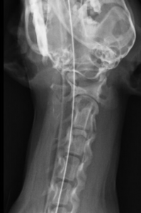
Dorsal view of a dog missing the dens of the axis
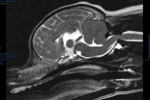
Sagittal view of a dog with a AA luxation seen on MRI
Treating atlanto-axial luxation?
Atlanto-axial luxation can be managed by two different ways:
- Conservative management. This involves cage rest and pain relief medication. Conservative treatment may need to be prolonged for several weeks, and although improvement can be initially expected in many patients, clinical signs often recur once the level of activity is increased again. If the reason for the AA luxation is a congenital malformation and not a trauma, this treatment is unlikely to give a satisfactory outcome.
- Surgery. The aim of the surgery is to permanently stabilize the atlanto-axial joint in a normal position, which reduces the neck pain and allows the spinal cord to recover. This procedure normally involves placing screws and bone cement or a special bone plate between the atlas and the axis to permanently stabilize the joint. Postoperatively, the patient will need 6-8 weeks of cage rest prior to returning to normal activity. The prognosis with surgery is generally good, but will vary depending on the dog’s age, severity of clinical signs, and degree of spinal cord damage. This is be a solution for the instability if your dog is born lacking the dens of the axis.


