Cerebrovascular Accidents
Back to Fact Sheets
Cerebrovascular accidents (CVA, stroke): owner information handout
Download PDF
What is a cerebrovascular accident?
A cerebrovascular accident (CVA), often referred to as a stroke, occurs when a part of the brain becomes dysfunctional due to a problem affecting its blood vessels. Strokes fall into two main categories- ischaemic and haemorrhagic. In an ischaemic stroke, an area of the brain is deprived of its blood supply due to an obstruction in the blood vessels. Haemorrhagic strokes result from a rupture of blood vessels. This type is less common in dogs and cats compared to ischaemic stroke.
As with all neurological diseases, the particular abnormalities that occur reflect the part of the brain that is affected and therefore a wide range of clinical signs can be seen. Loss of balance and coordination, abnormal posture, tilting of the head and flickering of the eyes may occur. Patients may find it difficult or be unable to walk. Disorientation, loss of vision, altered mental status and seizures are also possible.
Any age and breed of dog or cat may be affected, but strokes are most commonly seen in middle aged and older patients, and some breeds, such as the Cavalier Kind Charles Spaniels and Greyhounds, seem to be predisposed.
How is a stroke diagnosed?
There are some features of the clinical history that may be suggestive of a stroke. For example, abnormalities often occur peracutely, (meaning very suddenly)- the animal is normal one minute then the next appears to be unwell. Following the initial event, there may be deterioration for up to a day or so, often followed by a gradual improvement over days to weeks.
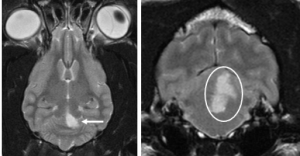
To make a confident diagnosis, MRI of the brain is usually recommended. In cases of ischaemic stroke, there are specific MRI sequences that can be used to help to diagnose the condition. Abnormalities can often be seen that very closely match an area of the brain supplied by a particular artery.
In the example below, the white approximately triangular area (arrowed and circled) is abnormal and corresponds to the part of the brain supplied by an artery called the rostral cerebellar artery, so we were able to diagnose an ischaemic stroke, very likely caused by a blockage of the rostral cerebellar artery.
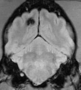
In cases of haemorrhagic stroke, a particular MRI sequence (termed gradient echo) is very useful for highlighting areas of haemorrhage (seen as the black arrowed area) on the image below:
Why has my pet had a stroke?
There are many potential underlying causes of stroke. Ischaemic strokes are caused by a small blockage within a blood vessel, and the blockage can be formed of a thrombus (ie a blood clot), abnormal cells (such as tumour cells), accumulations of bacteria and immune cells or even parasites. A thrombus can be caused by any disease process that alters blood flow, alters the composition of the blood, or increases the blood’s tendency to clot. Diseases that may predispose to clot formation include hypothyroidism, hypertension, diabetes, heart disease, kidney disease, overactive adrenal glands and tumours.
Tumours of many types also have the potential to cause a blockage if an accumulation of abnormal cells forms within a blood vessel. Infections can lead to a blockage as clumps of bacteria and immune cells may make their way into the bloodstream.
Some of the diseases mentioned above will show evidence on physical examination, and others can be screened for relatively easily with routine blood and urine tests. Blood pressure measurement can also be quickly and easily performed. It is likely that chest and abdominal imaging, usually by radiographs (x-rays) and/or ultrasound will be recommended in stroke patients in order to look for some of the potentially predisposing diseases that will not show up on blood tests and may not be obvious on physical examination, such as some types of cancer or infections.
The less common haemorrhagic stroke can be the result of diseases that stop the blood from clotting, toxins that inhibit clotting, liver disease, migration of parasites and tumours. Specialised blood tests can be performed that determine whether the blood is able to clot normally.
Around half of stroke patients have no identifiable predisposing factors. It is presumed in these cases that thrombus formation is responsible, but it is unknown why this occurs in some animals without any obvious trigger.
What treatment is required?
If a specific underlying disease is identified, treatment for that condition will be recommended. Sometimes, the MRI will show evidence of swelling of the brain and medications may be administered to address this. In cases of haemorrhagic stroke, bleeding may have led to the formation of a large clot within the tissues of the brain. Surgical removal of this may be required, though this will vary on a case-by-case basis and according to the suspected underlying cause.
If no underlying trigger for the stroke is identified, supportive treatment is administered while the animal recovers. This may include nursing care such as turning of the patient to prevent sores, bladder management, fluid and nutritional support and physiotherapy.
What is the prognosis?
The prognosis is affected by the presence of any underlying disease, and how easily this can be treated. In cases where no underlying disease is present, affected animals can make a very substantial improvement in a matter of days to weeks, even if they were initially affected by very severe neurological deficits. Some animals will show ongoing neurological abnormalities, but in many cases, this will not make a substantial impact on their quality of life.


