Chiari-like Malformation
and Syringomyelia
Back to Fact Sheets
The neurology department at Southfields provides a multidisciplinary approach to control neuropathic pain in dogs and cats suffering from Chiari-like malformation. We perform the current proposed caudal occipital malformation surgery as well as de-tethering surgical techniques popular in human patients. We work together with our anaesthesia service to offer pharmacological and non pharmacological options to improve the quality of life of our patients.
Download PDF
What is Chiari-like malformation and syringomyelia?
Chiari-like malformation (CM), and secondary syringomyelia (SM), is a painful inherited disorder common in Cavalier King Charles Spaniels but also possible in other brachycephalic toy breed dogs and cats.
CM is a complex developmental malformation of the skull and craniocervical vertebrae characterized by a conformational change, overcrowding of the brain, and subsequent partial herniation of the cerebellum through the foramen magnum. This can result in obstruction of the cerebrospinal fluid (CSF) channels at the craniocervical junction causing SM.
Syringomyelia is defined as a fluid-filled cavity within the parenchyma of the spinal cord. Its pathogenesis is poorly understood, but it is hypothesized to be extracellular fluid accumulation secondary to abnormal flow of CSF. SM can be secondary to CM and other pathologies that will alter the CSF flow.
How can you know your dog has Chiari malformation and/or Syringomyelia ?
CM/SM has a potential to affect your dog’s welfare due to chronic pain. However, certain patients with either CM or syringomyelia can be asymptomatic.
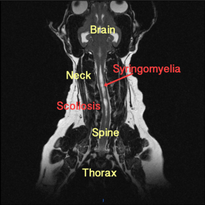
Clinical signs: Pain is the most distressing and common clinical presentation in animals developing SM secondary to CLM. Classical clinical signs of this type of pain include phantom scratching (which is a tendency to scratch towards one shoulder or neck region without skin), vocalization, scratching or rubbing of the facial region or ears, aversion to touch, refusal /difficulty jumping or going downstairs, and sleep disruption. Dogs with symptomatic SM may also present with neurological deficits such as scoliosis, weakness and difficulties walking.
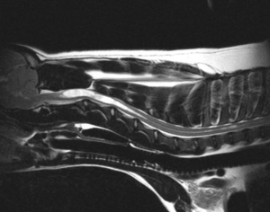
The standard of care for the diagnosis of CM/SM is ruling out other diseases that could mimic the clinical presentation. And the definitive diagnosis is achieved via MRI of the brain and the spinal cord.
In patients with CM, MRI shows caudal cerebellar herniation and compression, occipital dysplasia, and attenuation of CSF flow in the cranial cervical region. Syringomyelia appears as a linear hyperintensity within the spinal cord. Other concurrent, related abnormalities may include ventriculomegaly, kinking of the medulla, atlanto-occipital overlap, and dorsal compression at C1-C2.
What are the treatment options for your dog?
The primary goal of treatment is to manage neuropathic pain and neurologic deficits through pharmaceutical or surgical approaches. The majority of the symptomatic dogs and cats with CM/SM will have a progression of their clinical signs despites medical treatment and surgery might be advised; however, most of the cases maintain a good quality of life
Medical: We tend to advice to the majority of the cases to initiate a medical treatment before considering any surgical intervention. Our main goal is to improve the pain which is the main complain on these cases.
The corn stone of pain medication is the use of drugs that treat neuropathic pain. Neuropathic pain is caused by disease affecting the somatosensory nervous system and it is associated with abnormal sensations called dysesthesia or pain from normally non-painful stimuli (allodynia). We will use drugs like Gabapentin, Pregabalin, Amantadine, or Amitriptyline.
Anti-inflammatory drugs are routinely added. The first line medication will be non-steroidal anti-inflammatory drug (NSAID) but on certain specific cases corticosteroids might be needed. We tend to avoid long-term use of corticosteroids due to the potential side effects.
At Southfields we have developed a multidisciplinary pain clinic service, where acupuncture and other alternative therapies could be considered in selected patients.
Surgical: Surgery may be a possibility to treat the condition and it’s reserved to cases that failed the medical treatment. The goal of surgery is to decompress the foramen, allowing the fluid to flow normally. The most common surgical management is cranial/cervical decompression (also described as foramen magnum or sub-occipital decompression). The principle of this surgery is to allow CSF flow by removing the bone at the back of the skull (supraoccipital bone) and part of the top of the first vertebrae. Cranial/cervical decompression surgery is successful in reducing pain and improving neurological deficits in approximately 80% of cases with many cases doing well long term. However, about 50% of patients have a recurrence of signs within 2 years and the syringomyelia does not resolve.
Because of this relatively low long-term success of this surgery, the neurology department at Southfields offers also an additional surgery that is commonly perform in humans which purpose is to release the tension within the end of the spinal cord. This surgery is called tethered cord surgery and it has been successful improving cases that failed COMS surgery.
If you’d like to discussed any of these surgical, medical and alternative therapies for your pet, please contact our neurology department.


