Hydrocephalus
Back to Fact Sheets
Hydrocephalus is the active distension of the brain's ventricular system.
Download PDF
Hydrocephalus is the active distension of the brain’s ventricular system due to inadequate movement of cerebrospinal fluid (CSF) from its place of production in the ventricular system to the place of absorption. Although the most common form is congenital, it can also occur secondary to ventricular system obstructions.
Hydrocephalus classification
CSF behavior in hydrocephalus is important for classification and understanding this is critical to clinical decisions:
- Internal hydrocephalus: when the CSF builds up inside the ventricular system. For example, in congenital hydrocephalus or, secondary to a tumor or meningitis
- External hydrocephalus: when there is communication between the cerebral ventricular system and sub-arachnoid space (usually due to a blockage of the 4th ventricle) for example in bacterial meningitis
- Communicating hydrocephalus that occurs when the flow of cerebrospinal fluid is blocked after exiting the ventricles into subarachnoid space. This form is called a communicator because CSF can still flow between ventricles that remain open. The reabsorption of this fluid is altered in arachnoid villlosities by infections or bleeding. In this type of hydrocephalus the dilation of the ventricular cavities of the brain is dilated ahead of the site of the obstruction. This can be seen in congenital hydrocephalus, but also in inflammations or tumors
- Non-communicating hydrocephalus,also called obstructive hydrocephalus. Occurs when the flow of CSF is blocked along one or more of the narrowed pathways that connect the ventricles. One of the most common causes of non-communicating hydrocephalus is aqueduct stenosis. This type of hydrocephalus can also be seen in cats with feline peritonitis
- Compensated hydrocephalus or ex vacuo is secondary to a lack of brain matter where CSF fills the space. This destruction of brain material can occur in the uterus or later, due to significant trauma or vascular injury. We observe this type of hydrocephalus usually in puppies with a difficult birth in which prolonged hypoxia or vascular injury cause the absence of a good part of cortical tissue(hydroanencephalus)
- Normote dense hydrocephalus resulting from progressive blockage of CSF drainage pathways. Thus the cerebral ventricles slowly increase in volume and thus compression and damage of chronic brain tissue occurs. Normotense hydrocephalus is named after the brain ventricles grow without increasing intracranial pressure. Congenital hydrocephalus can be normotense and this is why the decision to treat surgical versus medical treatment can be difficult
- Hypertensive hydrocephalus occurs acute obstruction of CSF circulation producing acute intracranial hypertension. If not resolved, compression and damage of brain tissue is also caused by dilation of the ventricular system. It is typically seen in tumors and inflammations, but also in congenital hydrocephalus.
Clinical presentation
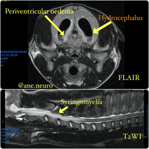
Signs directly related to hydrocephalus are secondary to alteration of intracranial pressure, secondary periventricular oedema (white matter), as well as loss in cortical neuron function (grey matter).
The breeds most commonly affected with congenital hydrocephalus are small and/or brachycephalic breeds (Maltese, Yorkshire Terrier, English Bulldog, Chihuahua, Lhasa Apso, Pomeranian, Poodle Toy, among others) but congenital hydrocephalus could also occur in large breeds even if it is less frequent.
The most common clinical signs are head enlargement or “dome shape” and ventro-lateral strabismus. Neurological signs demonstrate supratentorial dysfunction: altered mental state and behavior (difficult to train puppy), blindness, seizures and circling are the most common. When hydrocephalus is severe and hypertensive, secondary syringomyelia may develop and complicate the case with a multifocal location: propioceptive ataxia, cervical pain and tetraparesis.

Diagnosis
The most reliable and extensive method for diagnosing hydrocephalus and its cause is magnetic resonance imaging (MRI).
MRI will allow us not only to observe the enlarged ventricular system, but we will be able to diagnose the presence of inflammation, tumor injuries or congenital abnormalities. It will allow us to study the state of the peri-ventricular white substance and cortical state (brain sulcis) getting a more advanced idea of the type of hydrocephalus we are treating.
Treatment
The ultimate hydrocephalus treatment is surgery. However, in certain cases a palliative medical treatment might be suggested.
- Medical treatment Is there a medical alternative for the treatment of hydrocephalus? The only effective medical treatment, in the case of seizures, is the use of antiepileptics. Obviously antiepileptic treatment has no positive effect on the production or absorption of CSF, but it will reduce the frequency and intensity of seizures. In some animals where no other clinical signs are observed and there is no neurological progression, this may be the only treatment with satisfactory results.
Medical treatments available to reduce CSF production are few and do not usually significantly improve clinical symptoms: corticosteroids (anti-inflammatory doses) and diuretics
- Surgical treatment: Surgical treatment is the only treatment that will stop clinical development, controlling ventricular augmentation and as a result of brain degeneration.
At Southfields we offer a surgery called: Ventriculo-peritoneal shunting, which allows the release of the brain pressure by bypassing the CSF from the ventricles into the abdomen via an engineer develop silicon tube and valve.
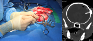
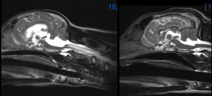
Before left and after surgery right
Our Head of Neurology Ane Uriarte performed the 1st of this kind surgery on a seal with hydrocephalus
https://www.nationalgeographic.com/news/2018/01/first-of-its-kind-brain-surgery-saves-beloved-fur-seal-spd/
Prognosis
If your pet is not badly affected and the hydrocephalus is discovered later in live the prognosis is generally good. These animals usually respond to antiepileptic treatment and generally do not usually need ventricular shunt.
In cases of rapid and hypertensive progressive hydrocephalus surgery is the only option, but in most cases the results are highly satisfactory. However, revision or replacement of the valve throughout the live of the animal is common.


